Molecular imaging can help to improve drug placement
Lucerne - Researchers from the University of Lucerne are working on a new molecular imaging technique. In this context, their aim is to use weakly radioactive tracers in conjunction with specialized tomographic techniques in order to deliver drugs precisely to a desired site of action.
Precision medicine is characterized, among other aspects, by patients receiving drug-based therapies in which specific substances are directed precisely to the site where their impact should be greatest. To this end, researchers from the University of Lucerne and the Hirslanden Klinik St. Anna, a hospital in Lucerne, are pursuing a new approach in which molecular imaging is used to identify the target structures for drug administration, as detailed in a statement.
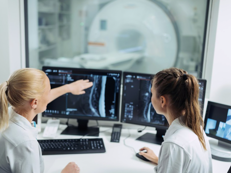
For this, they are using positron emission tomography (PET), among other techniques. The process involves the use of weakly radioactive substances – known as tracers – that bind to specific molecules in the body. These molecules, i.e. the drugs, can be precisely visualized on tomographic images, thereby ensuring that they reach their intended site of action.
In their study, the research team headed up by Martin Walter, Titular Professor of clinical-medical sciences and Head of Nuclear Medicine Research and Development at Hirslanden Klinik St. Anna, evaluated information on more than 1,600 approved drugs, over 700 therapeutic targets, more than 4,000 molecular imaging tracers and gene activity data from over 240,000 patients.
According to the analysis, approximately half of the target structures can be visualized. “Our long-term vision is to make molecular imaging an integral part of precision medicine”, as Martin Walter explains in the statement, adding that: “Rather than relying solely on tissue samples or genetic analyses, imaging can show where therapeutic targets are located throughout the entire body and how they change during treatment”.